
A synovial joint functions to unite long bones and to enable movement between them. They are characterised by having a fluid filled cavity which enables pain free articulation at the point of contact of the bones.
Synovial joints contain the following important structures
- Synovial cavity filled with synovial fluid
- Synovial membrane and joint capsule
- Articular cartilage
- Sub-chondral bone
2.1 Articular Cartilage
This is formed of hyaline cartilage which lines the epiphyses at the end of each long bone and provides the two articulating surfaces of the joint. It has a low number of specialised cells, chondrocytes, which act to maintain the cartilage matrix. This matrix is formed of collagen fibres, aggrecan and water with each critical to the function of the cartilage. It has no nerve or blood supply but derives nutrition from synovial fluid.
The cartilage must perform two critical functions
- Ensure smooth frictionless movement of the joint
- Sustain load during weight bearing
Cartilage matrix formation and destruction is crucial to normal function of a joint. The matrix is composed of proteoglycans, water and collagen. Proteoglycans are large complex molecules which resemble a test-tube brush. These are constructed from many sub-units or monomers which are arrays of glycosaminoglycans (chondroitin and keratin sulphate) attached to a central protein core. These units are bound to long hyaluronic acid (HA) chains to form large aggregates termed aggrecans. These provide two critical properties for articular cartilage. Firstly, the monomers are all negatively charged and so the whole array is trying to become as large in volume as possible because of the sidechains repelling each other. The other is that the array is hydrophilic, attracting and retaining water molecules. This whole arrangement can be thought of as the resilient or elastic part of the matrix which also acts like a sponge being able to be compressed and then returning to its normal form. The aggrecan arrays are restricted by a strong regular collagen matrix which imparts structure and strength to the matrix. When loaded the cartilage can be squeezed like a sponge with water content being pushed into the joint. When load is removed the cartilage resumes its shape by pulling water back into its substance. The collagen framework must allow this change in shape without being damaged.
Chondrocytes spaced evenly throughout the cartilage control the production and removal of matrix as well as its assembly to very exacting rules. In this way cartilage matrix construction and destruction are balanced. Chondrocyte activity is low in normal cartilage.
The load bearing and shock absorbing properties of the joint are further enhanced by the specialised structure of the sub-chondral bone supporting the cartilage.
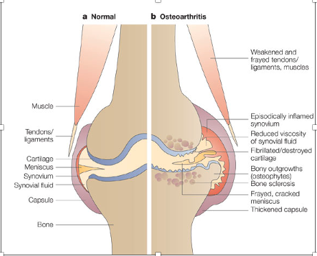
In osteoarthritic cartilage this balance is lost with matrix destruction predominating. Breakdown of the collagen structure allows the proteoglycans to expand by taking in more water. This is why cartilage in arthritic joints has a higher water content than in normal joints. Expansion of the proteoglycans coupled with load further disrupts the collagen skeleton and the physical tension of the cartilage is lost. This cycle continues with destruction of the complex framework within the cartilage. Changes within the sub-chondral bone with initial sclerosis and then microfractures accelerate damage by further reducing the cartilage’s capacity to withstand load.
2.2 The Synovium
The synovium comprises two components
- Synovial Membrane forming the inner layer
- Fibrous Joint capsule formed from the outer layer
- The Synovial Membrane
The synovial membrane is not a true membrane but acts as a sieve due to the hyaluronic molecule matrix and consists of:
- Intima – inner layer facing into the joint cavity containing synoviocytes
– 2 types of synoviocytes- Type A – macrophages; Type B – fibroblasts. It is typically 1-4 cells thick - Subintima – outer layer joined to fibrous portion of the joint capsule
contains connective tissue, blood vessels, lymphatic vessels and nerve fibres
Function
- The synovium acts as a semipermeable membrane to allow flow of certain particles in and out of the joint and maintains the composition of the synovial fluid through type A and type B intima cells.
- Type A cells – eliminate pathogens and excess material from the joint
– secrete cytokines and chemokines - Type B cells – Dominant cell in the healthy synovium
– Produce hyaluronic acid and lubricin
- Type A cells – eliminate pathogens and excess material from the joint
- The synovium in a normal joint has a low number of pain nociceptors
The synovial fluid is important in providing joint lubrication and nutrition for chondrocytes in the articular cartilage.
- The Fibrous Joint Capsule
This is the outer layer of the synovium, and it attaches to the periosteum of the long bones adjacent to the joint. It contains the joint ligaments and provides mechanical stability for the joint during movement. It must have a degree of elasticity to stretch when the joint moves into full extension or flexion. It is invested with many mechanoreceptors and nociceptors particularly around the point of ligament attachments. These act to prevent disruption of the joint by identifying extreme movement that could result in damage.
- Synovial Fluid
Synovial fluid is produced as a ultrafiltrate of blood plasma augmented by hyaluronic acid and lubricin by the Type B synoviocytes. It has a low cell count comprising mainly of macrophage type cells. It has three main functions namely
- To supply nutrition and remove waste from articular cartilage
- To lubricate the joint surfaces (cartilage and synovial membrane)
- Acts as a shock absorber when the joint is loaded in weight bearing
2.3 Progression in Pathophysiology in OA
Generally, the prevalence and severity of synovitis increases as the severity of OA increases and is associated with increased pain and reduced joint function.
Histologically, synovitis is characterised by synovial hyperplasia, fibrosis, neovascularisation and infiltration of macrophages and lymphocytes.
Activated synovial macrophages play an important role in OA through:
– production of pro-inflammatory cytokines (IL-1; IL-6; TNF -Alpha) and enzymes (MMPs) leading to articular matrix degradation and progression of the synovial inflammation.
– osteophyte formation
– activation of the complement cascade.
The fibrous joint capsule becomes thickened and less flexible.
Synovial fluid becomes less viscous, and both its lubricating and shock absorbing abilities are reduced. It can increase in volume and distend the joint capsule causing pain. Delivery of nutrients and removal of waste are both impaired. There will be more inflammatory components driving catabolic pathways and causing pain through activation of pain receptors within the joint. However, the cell count, and population are similar to that found in a normal joint.
To summarise, synovitis can result in lower concentrations of cartilage-protecting factors and high concentrations of factors that destroy cartilage. It can also reduce the capacity of synovial fluid to provide nutrition to the chondrocytes as well as reducing its lubricating and shock absorbing properties. Inflammation can also result in increased numbers and sensitivity of nociceptors within the synovium.
FIG 1 Intra-articular Pathology in OA
References
- Scanzello CR, Goldring SR. The role of synovitis in osteoarthritis pathogenesis. Bone. 2012; 51(2):249-57
- Monemdjou R, Fahmi H, Kapoor M. Synovium in the pathophysiology of osteoarthritis. Therapy 2010; 7(6): 661–668
- Hui AY, McCarty WJ, Masuda K, Firestein GS, Sah RL. A systems biology approach to synovial joint lubrication in health, injury, and disease. Wiley Interdiscip Rev Syst Biol Med. 2012; 4(1): 15–37.
- Kapoor M, Martel-Pelletier J, Lajeunesse D, Pelletier JP, Fahmi H. Role of proinflammatory cytokines in the pathophysiology of osteoarthritis. Nat Rev Rheumatol. 2011; 7:33–42
- Mazieres B, Garnero P, Gueguen A, Abbal M, Berdah L, Lequesne M, Nguyen M, Salles JP, Vignon E, Dougados M. Molecular markers of cartilage breakdown and synovitis at baseline as predictors of structural progression of hip osteoarthritis. The ECHODIAH Cohort. Ann Rheum Dis. 2006; 65: 354–9.
- Goldberg RL, Huff JP, Lenz ME, Glickman P, Katz R, Thonar EJ. Elevated plasma levels of hyaluronate in patients with osteoarthritis and rheumatoid arthritis. Arthritis Rheum. 1991;34:799–807