
Before considering how osteoarthritis acts to impair joint function it is useful to consider exactly what enables joints to function normally and the roles played by all the different tissues in ensuring normal pain free movement. When a macro view is taken it is surprising how complex this. This is a departure from the traditional focus on individual tissue pathology, which often dominates OA discussion. By understanding how the various tissues contribute and combine actions to ensure function it is easier to identify what is required to counter problems produced by a chronic progressive condition like osteoarthritis1. Osteoarthritis is a disease which is clinically defined by pain, but it is important to remember that pain is a sign caused by underlying and on-going pathology. Control of pain is fundamental to good disease management, but some attention must also be paid to the pathologies driving pain2,3. Removing pain without addressing this underling progressive deterioration of the joint structures only partly addresses the problems caused by the disease.
Synovial joints in normal animals allow flexible and effective movement. To do this they must withstand forces generated on the limb with every stride without becoming damaged. These forces can be recognised as load and forces generated by movement on the joint issues. A friction free joint surface is essential to prevent wear resulting from continual movement.
Articular cartilage, as an efficient composite tissue, can withstand loads of up to eight times bodyweight and has a friction co-efficient three times lower than ice on ice4 .
The body also has on-going capacity to repair any minor damage at source and restore efficient function. This capacity is depleted in osteoarthritic joints where damage exceeds the capacity for complete repair, and a downward spiral of continued damage and ineffective repair ensues resulting in the pain and loss of function that we see in the clinical disease.
The joint achieves its function by having the following
- Specialised tissue structures within the joint to allow shock absorption and friction free movement.
- Structural stability allowing free movement within well-defined limits allowed by the joint capsule; ligaments and tendons and controlled by a protective sensory system.
- Support and protection from the surrounding muscular elements coordinated by a finely tuned neuromuscular control system.
- Balanced biological system to sustain tissue function and allow repair.
In disease all these systems can become disrupted as the disease progresses. A degree of compensation can protect function up to a limit and then this too is overcome leading to failure of the joint as an organ1. Our purpose in managing the disease is to try and halt or limit some of the key disruptions leading to pain and continued pathology and at the same time encourage the healing mechanisms to predominate and restore better function.
Table 1 Failure of an Individual Joint
| Function | Tissue | Failure |
| Load Bearing | Cartilage
Sub-Chondral Bone Synovial Fluid Fibrous Joint capsule Tendons and Ligaments Muscles |
-Disrupted collagen architecture
-Loss of conformability -Structural disruption -Sclerosis -Loss of compressibility and protection of overlying cartilage -Decreased viscosity and loss of visco-elastic load sharing -Stretched allowing abnormal movement -Thickened and fibrous restricting movement by loss of elasticity and increased pain sensitivity -Stretched or torn allowing abnormal movement and load -Weakened increasing disruptive forces on joints -Poorly coordinated function due to reduced proprioception |
| Joint Movement | Cartilage
Synovial Fluid Synovial Membrane Fibrous Joint Capsule Muscles and tendons |
-Surface damage due to fragmentation and loss of surface lubrication system
-Loss of viscosity and lubricating ability leading to damage to articular surface and synovial membrane -Thickened impairing movement and becoming trapped resulting in further inflammation and pain – becomes thickened and loses compliance resulting in physical restriction of range of movement -Disuse atrophy causing weakness -Uncoordinated action causing inefficient movement -Compensation for other disruptions within the closest joint or other affected joints elsewhere |
| Biological Activity | Cartilage
Sub-chondral bone Synovial Membrane Peripheral and Central Nervous Systems Muscles and Tendons |
-Activated Chondrocytes with dysfunction and catabolism exceeding anabolism
-Release of pro-inflammatory cytokines and degradative enzymes -Chondrocyte senescence -Remodelling with increased osteoclast function -Vascularisation of the tidemark zone with cartilage -Neuro elements formed leading to sensitive articular surface when mechanical stress applied -Formation of Osteophytes and changes in bone shape due to osteoblastic activity -Synovitis with disrupted function with increased pro-inflammatory cytokines and degradative enzymes -Poor synovial fluid quality with consequences to mechanical function and poor nutrition supply to chondrocytes -Increased nociceptive elements leading to peripheral sensitisation Alteration in neuronal functions leading to both Peripheral and Central Sensitisation and Chronic Pain Degenerative changes in ligaments and peri-articular muscles |
Is there a recognisable sequence of progressive damage?
An affected joint will display different degrees of damage in many of these areas. This will alter depending on certain variables as will the clinical impact. Factors such as the joint involved, single, or multiple joint pathology, the existence of comorbidities and the individual’s response to pain will vary the presentation and the clinical problem. As the disease progresses with time the mix of pathological features will also alter. There is not a set sequence that the pathological changes follow over time but there is a general progression from the intra-articular events to those involving extra-articular structures. For clinical management purposes it is useful to identify three general regions of pathological changes
- Intra-articular Pathology
- Pathology of the nervous system
- Pathology of Disability; involving the supporting musculoskeletal tissues
Fig 2 Pathology extends beyond the joint to include neurological changes in both the peripheral and central nervous systems and the adjacent musculature
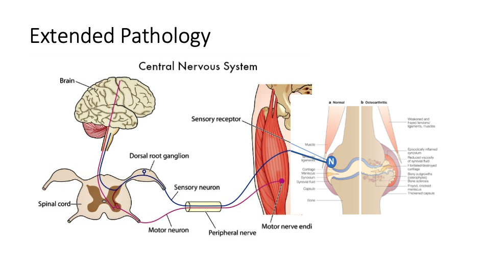
It is very important to recognise this and try to identify which of the pathologies is driving the disease at a particular point in time, and if there are any measures that can be used to specifically address this but targeting one or more of these areas.
References
- Loeser RF et al (2012) Osteoarthritis-A Disease of the Joint as an Organ. Arthritis and Rheumatism 64;1697-1707
- Hugle T and Guerts J (2017) What drives osteoarthritis? – synovial versus subchondral bone pathology. Rheumatology 56; 1461-1471
- Mathiessen A and Conaghan PG (2017) Synovitis in osteoarthritis: current understanding with therapeutic implications. Arthritis Research and Therapy 19; 18
- Eschweiler J et al (2021) The Biomechanics of Cartilage- An Overview. Life 2021, 11,302