
It is now accepted that pain is a key factor in OA. It is the reason for most owners with animals affected by osteoarthritis seeking veterinary attention. Knowing that pain is a major feature of the disease and that OA is the most common source of chronic pain in small animals makes a compelling case for better management.
Pain is essentially a sign of OA, but it also results in additional pathology which must be addressed in disease management. Chronic pain becomes part of the disease rather than a mere sign. We need to understand more about the disease and how it produces pain to allow us to better control this but also to aim to remove or reduce the cause of the pain in the first instance.
A synovial joint is a complex structure. It must fix in position during weight bearing but also must move freely to enable movement. These functions are made possible by its complex anatomy and physiology and must be controlled by a well-coordinated neuromuscular effort to prevent joint disruption.
Osteoarthritis represents the failure of the synovial joint as an organ1. But what about the pain that dominates clinical signs? In a normal joint, physiological pain acts to protect the joint from extreme ranges of movement with the potential to disrupt the supporting soft tissues. However, in osteoarthritis pain is pathological, recurrent and/or persistent. This chronic pain has no protective function and becomes part of the disease2, so called mal-adaptive pain. This chronic pain is characterised by the development of sensitisation of the nervous system both peripheral and central. The important result of this change is to reduce the actual level of stimulus required to evoke a significant response resulting in increased perception of pain3.
Fig 3 Complex interactions involving the various structures within the joint develop as the disease progresses and contribute to the complicated clinical picture
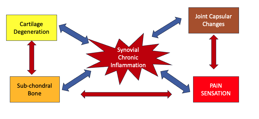
4.1 Pain perception in OA – ‘The problem is in the joint, but the Pain is in the Brain’
If we consider how pain is generated and perceived, the whole process is just a set of signals until it is processed centrally within the brain to produce the sensation we understand as pain. Stimulation at the periphery is detected by nociceptive neurones and conducted as action potentials to synapse with neurones comprising the spinal columns. There are two main types of afferent neurones defined by their myelination and conduction abilities, A and C fibre types. A-fibres (Proprioceptors) are thought to be responsible for proprioception and protective acute pain response signals, being larger diameter, well myelinated and exhibiting fast and precise action potential transport. C-fibres (Nociceptors) are thinner poorly myelinated and are believed to be responsible for the vaguer pain response characteristic of chronic pain2. The many stimuli arriving at this first base camp, the interaction between the primary peripheral and secondary spinal neurones, are modified and often amplified before passing to the spinal transport systems. Once again during this transit signals are modified by the actions of facilitatory or inhibitory inter-neurones to produce the final picture presented to various centres in the brain4. Note that right up to this point this is just a set of electrical impulses. The areas it is delivered to centrally as well as the magnitude of the stimulus dictates the subsequent pain response in terms of perception of site and impact on emotional and behavioural response2,3,4.
It is fair to state at this point that we still do not fully understand this process and why it remains somewhat unpredictable with different responses seen in different individuals.
In this way, osteoarthritis offers a perfect storm and real challenge for pain control. This is as a result of a number of disease features
- Persistent low-grade cartilage and synovial pathology results in constant stimulation of the joint nociceptive neurones
- Inflammation in the synovial membrane and the dorsal route ganglia provide additional sources of significant and constant stimulation.
- Alterations in the physical nature of the synovial membrane, particularly in the outer joint capsule and ligaments, reduce elasticity necessary to allow joint movement without tissue damage. Reduction in flexibility results in increased non-elastic stretch and potential tissue damage stimulating a protective nociceptive pain response.
- The complexity of the neural networks to enable joint function, present a large target for pathological malfunction especially when normal function and movement produces so much neural activity.
- Sensitisation of the peripheral system in the form of hyperalgesia and allodynia results, meaning minimal stimulation will evoke a significant signal to be passed to the spinal transport systems.
- This is further altered by plasticity of the neuro-matrix centrally which further distorts the sensory input (central sensitisation) and again making it more likely that incoming signals will be perceived as above threshold and evoke a pain response.
- The central neuro-matrix acts as a common pathway to all incoming traffic. Further stimulation from other sites will elevate response. OA is often present in multiple joints, each providing its own stimulation.
- Pain picture will change with advancing disease stage.
Chronic osteoarthritis is therefore typified by a pain picture which does not seem to be in concert with the contemporary pathology. Furthermore, as actual pathological changes result both in the peripheral nervous system and in the central neuro-matrix, these changes should be considered part of the essential pathology of the chronic disease.
Currently chronic pain in OA is still considered to be predominately driven by nociceptive pain and this conditions management efforts to try and reduce stimulatory inputs to wind-down sensitised neuro systems. This does take time, at least 90 days in an uncomplicated situation5. This explains the drive towards more long-term analgesic treatments. There is evidence that central sensitisation can be removed or abolished by removing nociceptive stimulation6. This has been demonstrated in cases of total joint replacement7
4.2 Sources of Pain in affected joints8
- Synovial inflammation
- Synovial effusion stretching joint capsule
- Inflexible joint capsule during joint movement
- Stretched periosteum over osteophytes
- Vascularised cartilage in advanced disease
- Microfractures in subchondral bone and potential medullary inflammation in medullary cavities and osteophytes
- Venous hypertension
- Muscular pain due to allodynia
- Muscle spasm due to disuse atrophy and damage during motion
- Joint instability due to failure of proprioceptive and coordinating neuromuscular systems
4.3 Pain influencing Clinical Presentations
When dogs or cats with OA show clinical signs, it is usually the result of pain. Hopefully the descriptions above show that this pain is very complicated and as such can manifest itself in different ways at different times and stages of the disease, appearing as overt pain producing obvious lameness or subtle behavioural changes. It is important to remember that many humans with pathological OA report no pain symptoms either at all or at time stages in the disease. It is also likely that different types of pain will show at different time points in the disease and often coexist. The resulting picture is that of a waxing and waning clinical presentation especially in the earlier stages which is best understood by being recorded over long periods of time rather than in single assessments. A single assessment may be very misleading about the actual impact of the disease if it is conducted at a particularly bad or good time in the clinical manifestation.
The challenge is for us to detect when pain, particularly chronic maladaptive pain is producing suffering in our OA patients and have an effective set of strategies to counter it.
References and Further Reading
- Loeser RF, Goldring SR, Scanzello CR and Goldring MB. (2012) Osteoarthritis – A Disease of the Joint as an Organ. Arthritis and Rheumatism 64; 1697-1707
- McDougall JJ (2019) Osteoarthritis is a neurological disease-a hypothesis. Osteoarthritis and Cartilage Open 100005
- Schaible H. Mechanisms of Chronic Pain in Osteoarthritis (2012) Curr Rheumatol Rep 14: 549-556
- Vincent TL and Miller RE (2024) Molecular pathogenesis of OA pain: Past, present and future. Osteoarthritis and Cartilage 32: 398-405
- Ley SJ, Livingston A, Waterman AE. The effect of chronic clinical pain on thermal and mechanical thresholds in sheep. Pain. 1989;39(3):353–357
- Van Greinsven H et al (2020) Central Sensitization in Musculoskeletal Pain: Lost in Translation? Journal of Orthopaedic and Sports Physical Therapy 50:592-596
- Arant K.R., Katz J.N and Neogi T (2022) Quantitative sensory testing: identifying pain characteristics in patients with osteoarthritis. Osteoarthritis and Cartilage 30 17-31
- Dye SF, Vaupel GI and Dye CC (1998) Conscious neurosensory mapping of the internal structures of the human knee without intraarticular anaesthesia. Am J Sports Med 26: 773-777